Children's Emergency Department is now located in Children's Tower: 1001 E. Marshall Street.
Learn more
Children's Emergency Department is now located in Children's Tower: 1001 E. Marshall Street.
Learn moreOne in 10 children is born with a vascular difference such as a hemangioma or vascular malformation. These are types of birthmarks caused by blood vessels that don’t develop correctly.
Our Center for Craniofacial Care has developed a comprehensive, multidisciplinary program to diagnose and treat individuals with all types of vascular differences. The physician specialists on our vascular birthmark team use the latest advances in medical, laser and surgical technologies to customize treatment plans for each child. Our team is skilled at treating a wide variety of conditions, including those listed below and related syndromes.
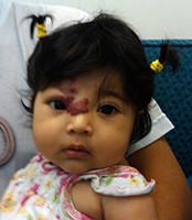
The most common vascular birthmark is a hemangioma, which is a type of benign (non-cancerous) growth found in infants. Hemangiomas are growths of blood vessel cells in a specific area and are also called vascular tumors. They are more common in girls.
Hemangiomas typically appear within a few days to weeks of an infant’s life and may look like a small red scratch or tiny bump. The hemangioma will grow quickly at this initial stage, which is known as the proliferative phase. The proliferative phase usually lasts six months to a year.
Most hemangiomas will then slowly get smaller without any medical treatment (called the involuting phase). During this time the color usually changes from bright red to a dull red and it may leave behind scarred or saggy skin. It can take as long as 10 years for this to happen. (In half of children this will happen by the time the child turns five and in most all, nearly 90 percent, by the time the child turns 9.) If the hemangioma is not significantly smaller and less noticeable by the time a child is of school age, treatment may be considered to reduce any effects on self-esteem.
Hemangiomas are a growth of abnormal endothelial cells in one area of the body. These growths are not cancerous. Endothelial cells are the cells that normally line the inside of blood vessels. Exactly why these growths occur is not known.
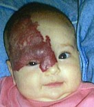
Hemangiomas look different depending on their location on the skin. If they are on the surface of the skin (superficial), they can have a bright red “strawberry” textured appearance. If they are under the skin (deep), they often look like a bruise or bluish swelling. A compound hemangioma is both deep and superficial.
While hemangiomas are not cancerous and usually do not cause any pain, they can lead to complications, including skin problems and bleeding. In some cases, hemangiomas can occur internally in areas such as the liver or airway and pose a health risk. These are called internal lesions.
Infants with three or more hemangiomas should have an ultrasound evaluation to look for any internal lesions. Large hemangiomas should prompt a search for other types of problems. They can be associated with vascular malformation syndromes which can have multiple related issues. Other testing such as MRI may be recommended.
If children with multiple hemangiomas or large hemangiomas are not growing and gaining weight, or are not reaching developmental milestones, they should be seen by a vascular birthmark team that includes specialists from different medical areas.
There are a few special types of hemangiomas which are different than described above. In some cases, a fully formed lesion is present at birth. These hemangiomas may not have a growth phase and may shrink rapidly or will never shrink and require surgery. Any hemangioma that does not follow the typical growth pattern should be evaluated by a multidisciplinary vascular birthmark team.
Hemangiomas may not require any treatment other than being monitored for growth. Sometimes medication may be recommended which may be applied directly to the site or taken by mouth to slow the growth. Medication, laser therapy and/or surgery may be recommended especially if there are any problems that affect a child’s function or issues with self-esteem.
Vascular malformations are different abnormalities that occur when blood vessels (veins, arteries, capillaries and lymph vessels) don’t develop properly before birth. Vascular malformations are considered a type of birthmark as they most often affect the skin, appearing as a red or blue color or swelling. They are entirely different from hemangiomas.
Vascular malformations can occur anywhere in the body. Typically, the malformation grows at the same rate a child’s body grows and may not be noticed for months or years, even though it is present at birth. This growth pattern differs from hemangiomas, which grow rapidly during infancy and then slow down and gradually shrink.
Often, vascular malformations cause little to no problems. Sometimes, however, this type of birthmark may start growing rapidly and out of proportion. This may happen because of some sort of injury to the area or things like going through puberty or pregnancy. If any type of vascular malformation is suspected, the child should be referred to a vascular birthmark specialist for evaluation.
Vascular malformations can be grouped depending on the type of vessel involved. These subgroups include: venous, arterial, lymphatic or capillary malformations or a combination of these types.
Venous malformations (VMs) are one of the most common vascular abnormalities. A VM is an abnormal formation, or mass, of tangled, enlarged veins somewhere within the body. On the skin, they can appear as a purple or bluish spot or just a bulge in the skin. These abnormal veins often lack smooth muscle along the vessel wall, causing them to enlarge and rupture, or burst, easily. The center of the tangled veins is called the nidus.
There are a few options for treating VMs depending on the size, location and type of symptoms. Usually, VMs are not treated unless there is a risk, such as a possible rupture or pressure on an organ, or if a child is having pain.
The most common methods of treatment are surgery and sclerotherapy. Complete removal of the nidus is required for an effective cure. Depending on the size and location of the mass, this can be very difficult, sometimes impossible, by surgery alone.
Sclerotherapy uses a chemical solution that is injected into the veins. The solution is very irritating to the walls of the veins and causes a clot to form. This cuts off the blood supply and the vein gets smaller. It usually takes several treatments before sclerotherapy shrinks the mass. It is done in an outpatient setting and does not require general anesthesia. There are no scars and the risks are low.
Arteriovenous malformations (AVMs) are abnormal connections between arteries (the blood vessels that carry blood away from heart) and veins (the blood vessels that carry blood back to heart). They are believed to be caused by the artery-capillary-vein connections forming improperly during early pregnancy. AVM’s may affect a specific area or be more widespread. They are often found on the trunk (torso) or on arms and legs.
The cause of most AVMs is not known. In childhood, the pink/red blush of color that appears on the skin with an AVM can be mistaken for a hemangioma. However, with a fast flow of blood-flow an AVM becomes noticeable. The skin may become a darker red or purple color, nearby veins may enlarge, a pulse may be felt in the area and the area may also feel warm.
When the amount of blood passing through the AVM is very large, the heart is affected because it has to handle more blood flow which can affect the heart’s functioning. AVMs are treated using techniques such as embolization (blood vessel blockage), sclerotherapy (shrinking of blood vessels) and surgery, or a combination of these techniques.
If the AVM occurs within the brain, a specialist in neurosurgery is required.
Lymphatic malformations are abnormal lymph vessels. These vessels are the tubes that deliver fluid from body tissues to veins as part of the body’s immune system. Malformations can occur when the fluid in the lymph vessels does not drain as it should. This causes excess fluid to build up creating cysts (fluid-filled sacs) or swollen, enlarged vessels. These larger vessels can then allow in more fluid, and sometimes even blood. Lymphatic malformations can occur in any part of the body but are most often found in the head and neck. If they occur close to the skin’s surface, they can resemble a bruise.
A lymphatic malformation may be seen on ultrasound before a baby is born. If a lymphatic malformation is suspected, it is critical the child is evaluated by a hospital vascular birthmark team who can work with the obstetrician and pediatrician to make sure the delivery is safe and the baby does well. Treatment usually involves sclerotherapy and/or surgery.
Capillary malformations (also known as port wine stains) are flat pink birthmarks that can darken and spread as a child grows. They are caused by enlarged capillaries (small blood vessels) in the skin and are often located on the head and neck, although they can appear in other areas of the body. They usually are not dangerous but will not fade on their own. In rare cases, they can be a sign of other conditions, so it’s important the child is evaluated by a vascular birthmark specialist. Laser therapy can be very helpful in children to lighten the area.
Each type of vascular malformation is treated differently and even two malformations of the same type need an individualized treatment plan depending on the size, location and symptoms. The treatment plan must be specialized for each child and usually requires coordination by a number of different specialists on our vascular birthmark team.